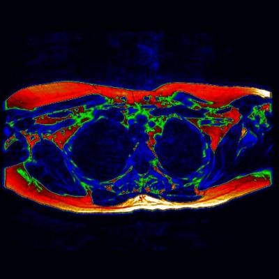
Identifying Brown Fat With Scans

Using a groundbreaking magnetic resonance imaging (MRI) technique, scientists are able to identify calorie burning "brown fat" for the first time in living adults, according to a recent study.
The study, published in The Journal of Clinical Endocrinology and Metabolism, suggests that studying how brown adipose tissue -- commonly called "brown fat" -- functions in the body will help scientists develop new and effective treatments for obesity and diabetes.
Brown adipose tissue (BAT) is the "better" of two types of far tissue long suspected to be present in humans. The other fat, white adipose tissue (WAT) is more commonly seen, and is created when the body stores excess caloric energy for later use.
According to past studies, the main purpose of BAT is to generate body heat in an attempt to preserve regular body temperatures when it is cold. However, to do this, the brown fat burns calories at a very high rate, helping maintain weight in people who tend to exceed their recommended daily caloric count. Unsurprisingly, this fat is found with greater prevalence in thin people, compared to heavy people.
But while scientists have long known about BAT, they have had little chance to study how it functions within the human body, as current technologies made it difficult to distinguish WAT from BAT. Even positron emission tomography (PET) scans, which have been used to identify BAT in the past, have severe limitations as they do not so-much show the fat itself as they show its activity.
However, researchers from the University of Warwick have now shown that a new novel MRI technique can be used to produce clear images of BAT, allowing researcher to examine exactly how the fat functions and even appears in the body.
According to the study, the research team successfully identified brown fat in a 25-year-old women who was undergoing an MRI. The results detailed where the fat can be found in adults, allowing for new questions to be asked regarding its formation and activation.
The study was published the January Issue of The Journal of Clinical Endocrinology and Metabolism.
Apr 17, 2014 05:46 PM EDT


